IFM belongs to the family of immunohistochemical analytical methods for the detection of proteins in the objects of cultural heritage. The general principles underlying immuno-fluorescence microscopy, or IFM, are similar to those of ELISA; however, in the case of IFM, detection is achieved using a fluorescence microscope and secondary antibodies that are conjugated to a fluorophore. In short, a sample is embedded in epoxy resin and polished with successively finer grades of micro-mesh abrasive cloths and a felt for final polishing. The exposed sample cross sections are then incubated in the primary antibody dilution, are washed, are subsequently incubated in the secondary antibody dilution, are again rinsed and dried. A layer containing a matching protein binder to the primary antibody will be identified by a fluorescence detector of a fluorescence microscope.
Fields of application
-
Cultural heritage
archaeological object and site, decorative arts, manuscript, musical instrument, painting, photo, sculpture
-
Natural heritage
animal product, botanic collection, fossil, object in formalin, shell, skeleton, taxidermy collection
Materials
-
organic
animal parts, binding media, glues, varnishes
TOOLS
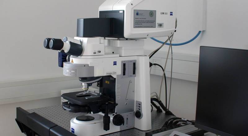
Zeiss confocal microscope LSM 800 MAT equipped with 405, 488, 561, 638 nm lasers allowing immunofluorescence materials characterisation and topographic analysis.